


Right Ventricular Infarction
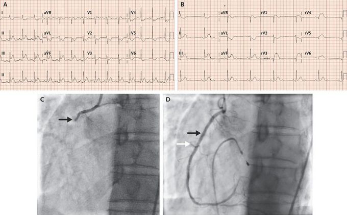
A 61-year-old man with a personal history of smoking and a family history of coronary artery disease presented with what he described as a squeezing pain in the left side of his chest that woke him from sleep. He also had associated dizziness and diaphoresis. His heart rate was 85 beats per minute, and he was hypotensive (blood pressure, 90/60 mm Hg). An initial electrocardiogram (ECG) showed a normal sinus rhythm, with ST-segment elevation in leads II, III, aVF, and V1 (Panel A). Right ventricular infarction was suspected because of ST-segment elevation in V1. ECG with precordial leads on the right side was performed and showed 2:1 atrioventricular block and ST-segment elevation in leads rV3 through rV6, which confirmed ST-segment elevation myocardial infarction of the inferior wall, with involvement of the right ventricle (Panel B). Intravenous fluids were administered, and a temporary transvenous pacing wire was placed. Coronary angiography revealed a right coronary artery with 100% occlusion proximal to the right ventricular branch (Panel C, arrow). Percutaneous coronary intervention was performed, and flow to the right coronary artery (Panel D, black arrow) and the right ventricular branch (Panel D, white arrow) was restored. With adequate fluid resuscitation and the restoration of flow in the blocked artery, the patient’s hemodynamic status improved, and the temporary pacing wire was removed the next day. Subsequent echocardiography revealed normal left ventricular systolic function and mild right ventricular dysfunction.
