


Cutaneous Actinomycosis
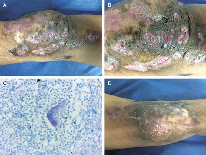
A 33-year-old man was referred to our institution in 2015 with lesions on his left knee that had worsened over the course of several years. In 2010, at which time he had an unremarkable medical history, he was involved in a motor vehicle collision that resulted in several superficial soft-tissue bruises on his left knee and no associated fractures. Two years after the collision, the patient sought medical attention for skin thickening and several subcutaneous nodules smaller than 1 cm that had gradually developed on the medial and lateral sides of his knee and had subsequently disseminated to the thigh. The patient reported no fevers or other systemic symptoms. He received several courses of empirical antibiotics and had minimal clinical improvement. On physical examination during the current presentation, the left knee was noted to have edema, erythema, hyperpigmentation, hyperkeratinization, and several indurated nodules with scabs and sinus tracts, but without purulence or granule secretion (Panels A and B). The patient had a normal range of motion and muscular strength, although there were some areas of hypoesthesia on the knee. He underwent surgery with débridement, and intraoperative cultures grew actinomyces species, a finding that was corroborated by histologic examination, on which an eosinophilic halo — known as the Splendore–Hoeppli phenomenon — was observed surrounding the organism (Panel C). He was treated with amoxicillin–clavulanic acid, administered orally for 1 year, and had clinical improvement but not full resolution (Panel D).
