


Squamous-Cell Carcinoma of the Tongue
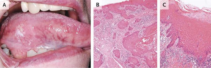
A 73-year-old woman presented with a 1-year history of a persistent ulcer and white patch on the right margin of the tongue and soreness while eating. She did not smoke or drink alcohol. Extraoral examination revealed no cervical lymphadenopathy. Intraoral examination revealed a well-circumscribed superficial ulcer, measuring 7 mm by 3 mm in the greatest dimensions, with a homogeneous base, an indurated upper border, and an adjacent speckled red and white patch at the lower border (Panel A). Incisional biopsy of the indurated upper border revealed invasive squamous-cell carcinoma (Panel B; hematoxylin and eosin). Incisional biopsy of the speckled area revealed hyperchromic and pleomorphic cells in the basal epithelial layer and a “budding” architecture, which was regarded as mild dysplasia (Panel C; hematoxylin and eosin). A computed tomographic scan confirmed no neck-node involvement. After wide excision of the carcinoma, the patient has remained free from recurrence for the past 12 months. The 5-year survival rate among patients with squamous-cell carcinoma of the tongue remains stubbornly low at approximately 50%, probably because of late diagnosis. The current case shows the value of multiple biopsy sites when the presentation is heterogeneous in appearance, as well as the importance of a high index of suspicion for carcinoma.
