Placenta Increta
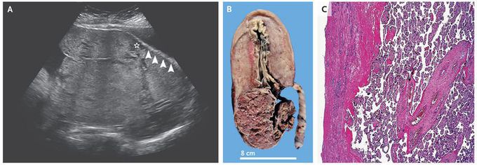
A 38-year-old woman who had undergone three low-segment cesarean sections was admitted to the hospital in labor at 36 weeks of gestation. Antenatal ultrasonography suggested placenta accreta (Panel A), with the appearance of irregular vascular spaces (asterisk) and loss of retroplacental hypoechoic stripe (arrowheads), which suggested obliteration of the basal decidua. She underwent an emergency cesarean section, which confirmed the abnormal adherence of the placenta to the myometrium. The placenta was left in situ, and a cesarean hysterectomy was performed to decrease the risk of massive hemorrhage. Gross and histopathological examination (Panels B and C) confirmed the diagnosis of placenta increta, with trophoblastic invasion into the myometrium. There is a spectrum of abnormal placentation: placenta accreta refers to the attachment of the chorionic villi to the myometrium, placenta increta to the invasion of the chorionic villi into the myometrium, and placenta percreta to invasion through the myometrium, with or without involvement of surrounding structures. Abnormal placentation may be suspected on ultrasonography or magnetic resonance imaging but can be diagnosed definitively only on histopathological examination. The patient’s postoperative course was uncomplicated. She was discharged home with her newborn on the fifth postoperative day, and both mother and child remained well at follow-up 12 months after delivery.