


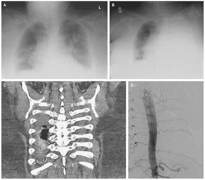
Hemothorax after Thoracentesis
A 65-year-old woman with atherosclerotic cardiovascular disease and renal failure was hospitalized for acute respiratory failure and bilateral pleural effusions associated with pneumonia (Panel A). To rule out the presence of empyema, a diagnostic, ultrasonography-guided thoracentesis was performed in the left lung in the 9th and 10th intercostal spaces. The initial fluid that was extracted had a brown discoloration but was transudative and was thought to be associated with the patient’s cardiorenal disease. Unfortunately, hemothorax developed shortly after the procedure (Panel B). Computed tomography and angiography of the chest revealed tortuous intercostal arteries, which had potentially contributed to iatrogenic arterial injury (Panel C). The hemothorax was managed successfully with chest-tube insertion and angiographic coil embolization (Panel D), and the patient’s clinical condition improved. There have been reported correlations between increased tortuosity of intercostal arteries and decreased safe space for thoracentesis, particularly in older patients.
