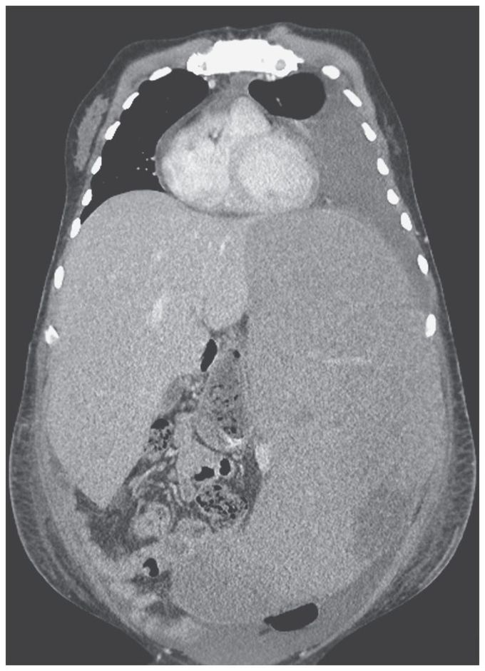


Massive Splenomegaly
A 35-year-old man presented to the emergency department with a 3-week history of fatigue and painful distention of the left side of his abdomen. He had a history of hepatosplenic T-cell lymphoma and had completed treatment 9 months earlier. Physical examination revealed massive enlargement of the liver and spleen, with the spleen crossing the midline and its lower margin extending into the pelvis. A computed tomographic scan of the abdomen confirmed hepatomegaly and splenomegaly (with the spleen measuring 36 cm in its greatest dimension). The white-cell count was 17,400 per cubic millimeter (reference range, 4000 to 11,000), the hemoglobin level 3.9 g per deciliter (reference range, 13.3 to 17.7), and the platelet count 10,000 per cubic millimeter (reference range, 150,000 to 450,000). A blood smear showed atypical mononuclear cells, intermediate to large in size, with irregular nuclear contours, moderately fine chromatin, prominent nucleoli, and some cytoplasmic blebs. Relapsed hepatosplenic T-cell lymphoma was confirmed on flow cytometry. Other causes of massive splenomegaly include chronic myeloid leukemia, myelofibrosis, hairy-cell leukemia, malaria, β-thalassemia major, and visceral leishmaniasis. Treatment with immune checkpoint blockade and opioid analgesics was initiated, but the patient died consequent to disease progression 10 weeks after presentation.
I dont understand the CT. Why does seem parient is enlarged posterior and anterior in the middle line? 🤔

