


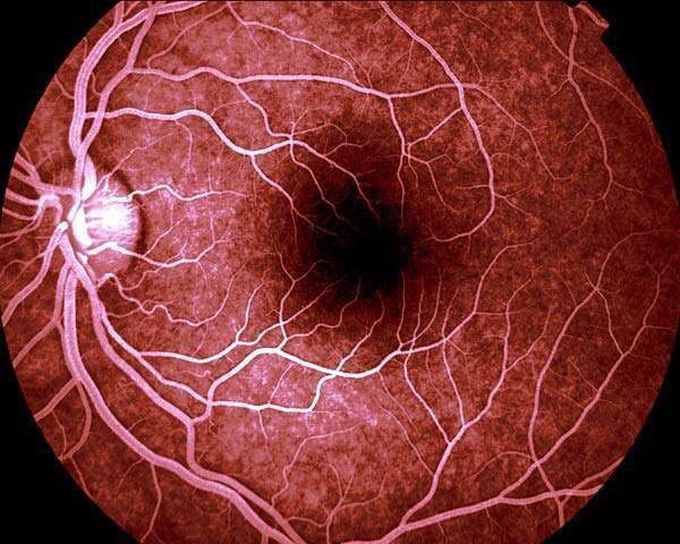
The back of our eye: Image of retinal funduscopy.This is what the doctor see when he examines your eye with the gadget that looks like a lamp with a magnifying glass :) Imagine you look through the pupil to the back of the eye, that's how it's going to look (or at least similarly). You can see the retina with the many supplying little arteries. They enter the eye together with the optic nerve -that comes from the brain- through the papilla nervi optici, the spot on the left. This is called "blind spot", because there are no photoreceptors on on that spot.The dark spot is the fovea centralis, the spot where your eye sees best (when you focus your eyes on an object, you project it towards the fovea centralis, because most of the photoreceptors are present here.
