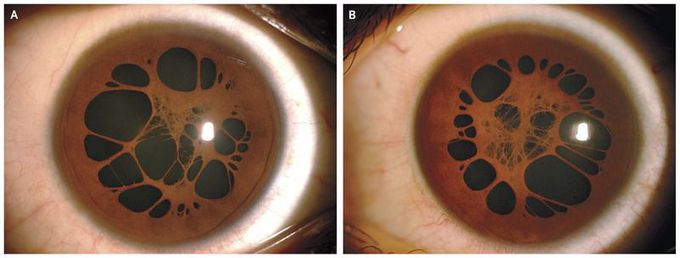
Persistent Pupillary Membrane
A 15-year-old boy presented with decreased vision in both eyes. His best corrected visual acuity, as determined with the use of the Snellen chart, was 20/20 in the right eye and 20/60 in the left eye, with anisometropic amblyopia in the left eye. A routine slit-lamp examination showed evidence of persistent pupillary membranes in both eyes (Panel A shows the right eye, and Panel B the left eye; see the. These membranes represent remnants of the tunica vasculosa lentis, which is the blood supply for the developing lens of the fetus. Remnants of the capillaries may persist as small strands attached to the collarette of the iris. Vision is usually unaffected, but occasionally thick, persistent pupillary membranes can cause deprivational amblyopia (i.e., stimulus deprivation to the retina because of any media opacity, leading to amblyopia), which can be managed with mydriatic agents, surgical excision, or laser-induced lysis. This patient was treated with application of a patch to the right eye for a few hours each day. At follow-up 3 months after presentation, visual acuity in his left eye had improved to 20/40.