


Grouped Pustules on an Erythematous Base
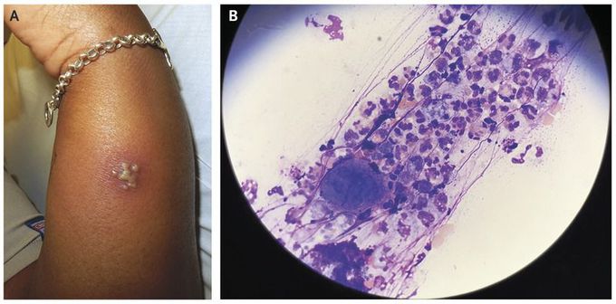
A 27-year-old woman with a history of genital herpes presented to the emergency department with painful, burning lesions on her left arm. On physical examination, grouped, coalescing pustules that had scalloped borders on an erythematous base and that were tender to palpation were seen on the left arm (Panel A). One of the pustules was unroofed (i.e., the top of the pustule was removed) and the base scraped, smeared onto a glass slide, and stained with Wright–Giemsa stain (Tzanck smear). Microscopic examination revealed numerous multinucleated giant cells with molded nuclei and marginated chromatin (Panel B), which together suggested a human herpesvirus infection. A polymerase-chain-reaction assay confirmed the presence of herpes simplex virus type 2. Morphologic features of clustered, coalescing pustules or vesicles on a red base are characteristic of herpesvirus infection. A Tzanck smear provides a rapid means of diagnosing herpes simplex infections and varicella–zoster virus infections. After treatment with valacyclovir at a dose of 1000 mg twice daily for 7 days, the patient had complete resolution of her symptoms and physical findings.
