


Melanoma of the Foot
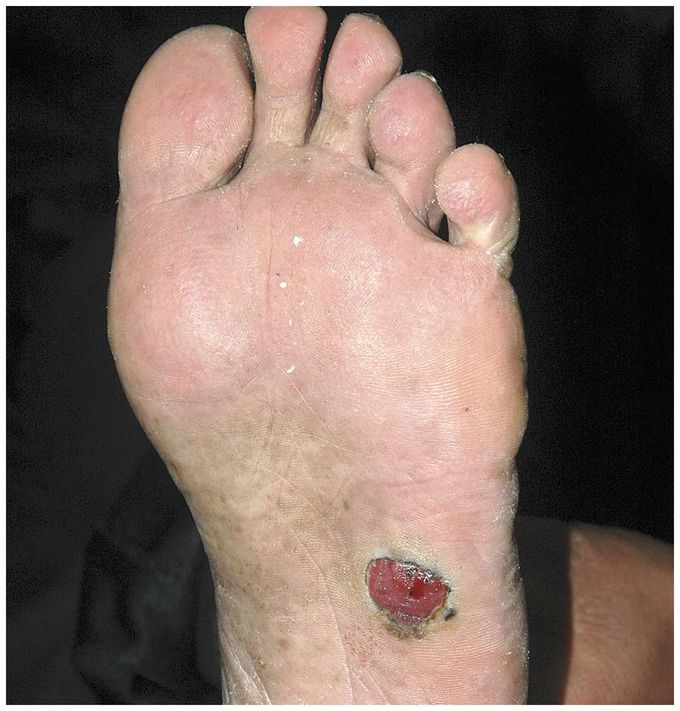
An 80-year-old man presented with a lesion on the left sole that had been increasing in size during the past 2 years. The lesion had become increasingly ulcerated, with intermittent bleeding. The physical examination revealed a well-defined, fleshy, reddish plaque measuring 2 cm in diameter with erosions on the surface and black areas at the periphery. A melanoma with a Breslow depth of 1.1 mm was identified on biopsy. Immunohistochemical examination showed tumor cells that were positive for S-100 protein and HMB-45 and negative for AE1/AE3. On the basis of these findings, a diagnosis of amelanotic melanoma was made. The general absence of pigment in amelanotic melanoma can lead to confusion with more benign skin conditions, including pyogenic granulomas, warts, and ulcers. Prompt investigation is important to avoid a delay in diagnosis. The patient was referred to an oncology clinic for further treatment but did not attend and was lost to follow-up
