


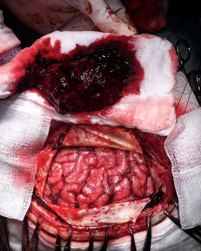
Huge left-sided spontaneous subdural hematoma in a 63-year-old male patient secondary to warfarin usage for his prosthetic cardiac valve.
This is a type of intracranial hemorrhage that occurs beneath the dura (over the surface of the brain). The patient presented to the ER with a Glasgow coma scale of 10, aphasia (comprehension of speech and the ability to read or write due to damage to the parts of the brain that control language), and right hemiplegia! A subdues hematoma occurs not only in patients with severe head injury but also in patients with less severe head injuries, particularly those who are elderly or receiving anticoagulants. In a chronic subdural hematoma, small veins on the outer surface of the brain may tear, causing a gradual and slow bleeding in the subdural space, which can hide symptoms for several days or weeks. Small subdural hematomas can be managed by careful monitoring until the body heals itself. Other small subdural hematomas can be managed by inserting a temporary small catheter through a hole drilled through the skull and sucking out the hematoma. The big ones require neurosurgical intervention. This picture shows the skull wide open with the brain completely exposed after performing a craniotomy, the dura mater incised and the hematoma being carefully evacuated.Photo credit @rpaglioli
