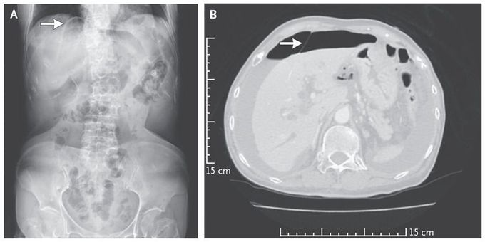


Falciform Ligament Sign
An 87-year-old woman with a history of hypertension and peptic ulcer disease presented to the emergency department with a 3-day history of abdominal distention, pain in the epigastrium, and watery diarrhea. She had hypotension (blood pressure of 78/49 mm Hg), and diffuse abdominal tenderness with guarding was observed on physical examination. An abdominal radiograph that was obtained with the patient in a supine position revealed the falciform ligament sign (Panel A, arrow) — a radiographic sign of pneumoperitoneum. The falciform ligament connects the liver to the anterior abdominal wall. When surrounded by intraperitoneal free air, the falciform ligament may be seen as a vertical band of soft tissue on a computed tomographic scan of the abdomen (Panel B, arrow). An emergency laparotomy was performed, and the diagnosis of a bowel perforation was confirmed; a 2-cm perforation in the duodenum was repaired. The patient was discharged from the hospital 22 days later and recovered well.

