


Medicaltalks over 7 years ago
#NSFWSensitive content 18+
This material contains content which some users might find distrubingPerinatal intracerebral hemorrhage
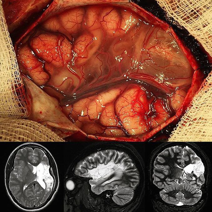
Here's a case of a 17 year old female patient that had a perinatal intracerebral hemorrhage, strictly means a pathologic accumulation of blood within the cranial vault. She then started having epileptic seizures at the age of 7.Over time they got worse and became refractory to medication in the last months. Imaging studies revealed a localized area of cerebral softening (encephalomalacia) which developed as a complication of the intracerebral hemorrhage. Neurosurgeons were able to resect the encephalomalacia in the perisylvian area which was causing the seizures. Below are the axial, sagittal, and coronal views of her MRI scan. Credit to @rpaglioli
Top rated comment
over 7 years ago
The imaging shown is mri taken how many days after hemorrhage?
Other commentsSign in to post comments. You don't have an account? Sign up now!
