


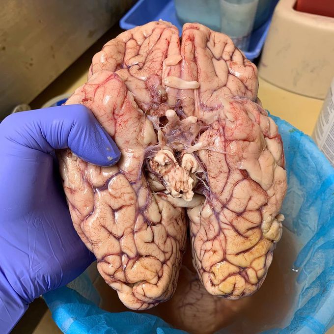
Bottom/inferior view of the brain!!
In this picture you can see some great things. Clearly you can see the optic nerve, optic chiasm, frontal and temporal lobes, and many others. The cerebellum is removed. These brain parts are marked with visible gross features like the gyri and sulci of the cerebrum and cerebellum. They are each also divided into subparts or regions for simplified localization of structures. For example, the brainstem is composed of the midbrain, pons and medulla oblongata, while the cerebrum is divisible into lobes. Sulci are small grooves; but there are also large grooves, and these are called fissures. Fissures divide the cerebral cortex into lobes, and also divide the cerebrum into the right and left cerebral hemispheres along the sagittal plane, thus partitioning the structure called the corpus callosum into two halves. Furthermore, the structures seen at this view are specifically referred to as the cortex, the most superficial layer of these brain parts, and make up the largest portion of the brains gray matter. Photo by @rustyyyjonesss
Hemodynamic stimuli&nonhemodynamic stimuliEffects of sugar on teeth
