
Metastatic Uveal Melanoma
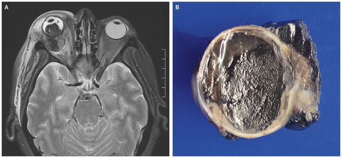
A 59-year-old woman presented to the emergency department with a 4-day history of inflammation and pain in the right eye. She had been blind in the eye for several years before presentation. The physical examination showed proptosis of the right eye, with periorbital inflammation, ophthalmoplegia, and a right relative afferent pupillary defect. Magnetic resonance imaging revealed a right orbital mass measuring 2.8 cm by 2.5 cm by 2.3 cm with intraocular and extraocular components (Panel A). The patient’s levels of alanine aminotransferase and aspartate aminotransferase were normal, but there were elevations in the alkaline phosphatase level (245 U per liter; reference range, 31 to 95) and the γ-glutamyltransferase level (225 U per liter; reference range, 7 to 37). Abdominal and thoracic imaging showed numerous hepatic masses, abdominal and thoracic lymphadenopathy, and vertebral sclerotic osseous disease, findings that were consistent with widely metastatic disease. The right eye was enucleated for palliative relief and to obtain tissue for diagnosis (Panel B). Immunohistochemical evaluation supported the diagnosis of uveal melanoma. The patient was treated with ipilimumab and nivolumab, but she died from progressive disease 2 months after presentation.
