


Temporary Ectopic Implantation of a Single Finger Using a Perforator as a Feeding Vessel, and Subsequent Prefabricated Chimeric Flap Transplantation
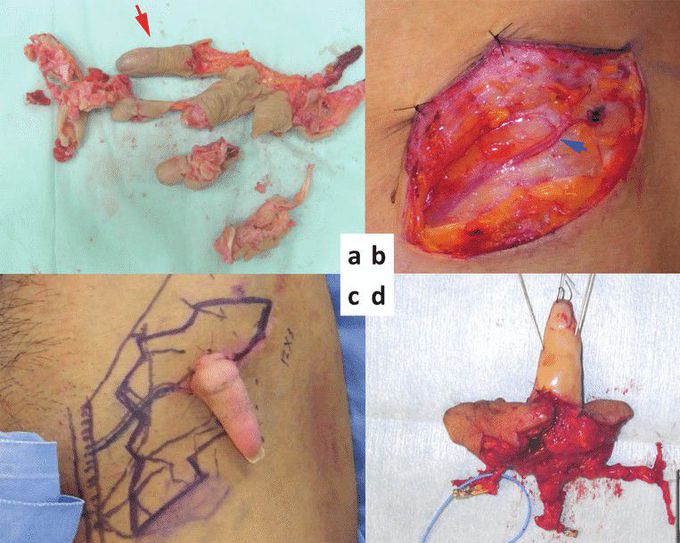
(a) Amputated little finger (arrow). (b) Left groin superficial circumflex iliac artery perforator (SCIP) (arrow). (c) Extopic chimeric flap design 3 months after ectopic implantation. (d) Chimeric SCIP flap with vascularized lateral femoral cutaneous nerve. Ectopic implantation was first reported by Godina, who implanted a subtotal amputated hand in the axilla in 1986.1 In 1996, Graf et al2 temporarily implanted ring and little fingers amputated at the level of metacarpophalangeal joints into the forearm. Salvage of amputated thumbs temporarily implanted into the ulnar and dorsalis pedis was reported by Li et al3 in 2008. We present 2 cases in which amputated fingers were salvaged and reconstructed by means of temporary ectopic implantation using perforator vessels and prefabricated chimeric flaps. CASE REPORTS Case 1 A 30-year-old man suffered a mangling injury of his right hand after it was caught in a meat grinder. All fingers were severely crushed with the exception of the little finger, which was intact distal to the proximal interphalangeal joint (Fig 1a). The hand was also crushed and ablated. We planned an ectopic implantation of the little finger to provide enough time to fully debride and optimize the hand in preparation for reimplantation of the little finger. The superficial circumflex iliac artery perforator (SCIP) and subdermal vein were first identifiednear the anterior superior iliac spine (Fig 1b). The diameter of the SCIP and the little finger digital artery were 0.4 mm and 0.6 mm, respectively. The digital artery of the little finger was anastomosed to the SCIP by using IVaS method.4 Three months after ectopic implantation of the little finger, the little finger was elevated with an SCIP flap (12 × 3 cm), containing the lateral femoral cutaneous nerve (4 cm), a superficial circumflex iliac artery and vein (SCIA and SCIV), and superficial inferior epigastric artery (SIEA) (Figs 1c and 1d). Because the litter finger is shorter than a normal size finger, the right second metacarpal bone with a dorsal metacarpal artery was harvested and placed distally on the 5th metacarpal bone (Figs 2a and 2b). The SCIA and SIEA originate from a common trunk, which was anastomosed to the base of the 5th common digital artery. The dorsal metacarpal artery of the second metacarpal bone was anastomosed to the distal end of the SIEA (Fig 2d). The SCIV and subdermal vein of SCIP flap were anastomosed to the dorsal veins of the hand. The vascularized lateral femoral cutaneous nerve was anastomosed to both ends of the 5th digital nerve. Sensation in the little finger recovered, with a Semmes-Weinstein test result of 4.08 and moving 2 PD of 8 mm at 10 months, and SCIP flap also had sensation with a Semmes-Weinstein test of 4.31.
