


J
JustSomeMedStudentabout 8 years ago
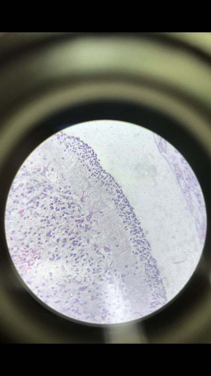
Look at those Purkinje cells!!
Cerebellar cortex under optic microscope, dyed with H&E stain. Notice the Purkinje cell in the centre of the picture, it’s actually possible to see how it branches out it’s dendrites.
Other commentsSign in to post comments. You don't have an account? Sign up now!
