
MRI Technologistabout 7 years ago

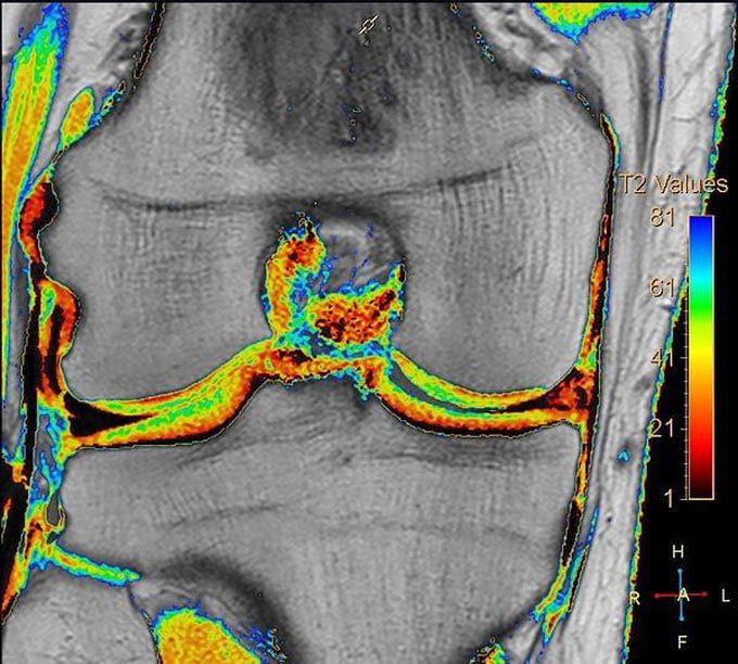
Figure shows T2 mapping of the knee articular cartilage, courtesy of Philips Medical Systems. Note that deeper layers near the bone have shorter T2 values (orange). Trauma, degeneration, and inflammatory changes in articular cartilage are generally manifest by increased free water content and loss of proteoglycans, causing increased T2 relaxation times.

