


Zunaira saleh10 months ago
MTA seal
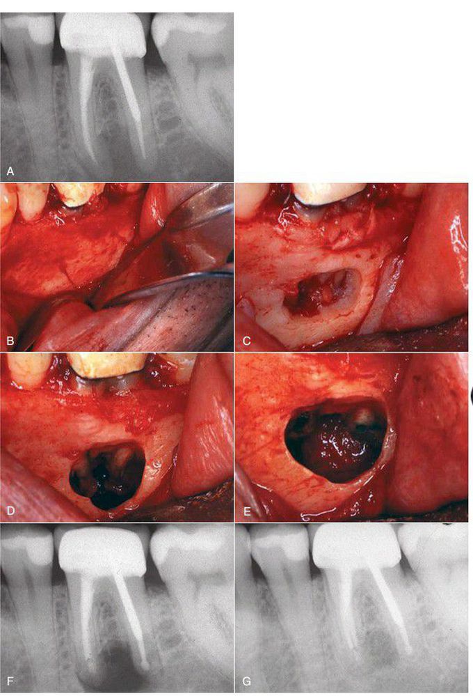
A preoperative radiograph showing the periapical pathologic condition amenable to apical surgery. (B) Full-thickness mucoperiosteal flap to expose lateral border of mandible. As is typical, no obvious bony perforation exists. (C) Careful removal of the thick buccal bone to expose the apical portion. (D) Apical one-third exposed before resection of root. (E) Both roots resected and mineral trioxide aggregate seal placed following ultrasonic preparation. (F) Immediate postoperative radiograph with mineral trioxide aggregate seal visible. (G) Five months after surgery, bone fill is evident.
Other commentsSign in to post comments. You don't have an account? Sign up now!
