
Zunaira salehover 1 year ago

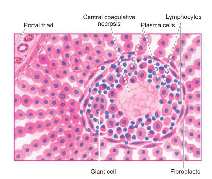
Syphilitic gumma
Typical microscopic appearance in the case of syphilitic gumma of the liver. Central coagulative necrosis is surrounded by palisades of macrophages and plasma cells marginated peripherally by fibroblasts.
Other commentsSign in to post comments. You don't have an account? Sign up now!

