
Zunaira salehover 1 year ago

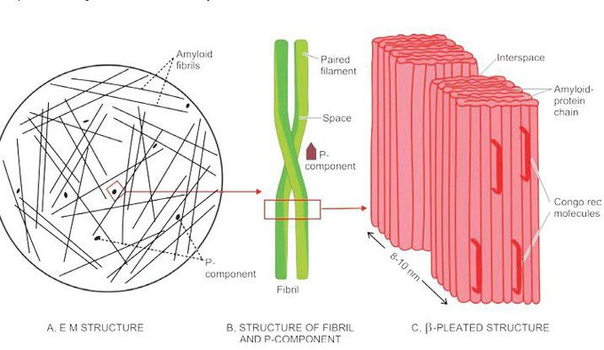
Diagrammatic representation of the ultrastructure of amyloid.a
A, Electron microscopy shows major part consisting of amyloid fibrils (95%) randomly oriented, while the minor part is essentially P-component (5%) B, Each fibril is further composed of double helix of two pleated sheets in the form of twin filaments separated by a clear space. P-component has a pentagonal or doughnut profile. C, X-ray crystallography and infra-red spectroscopy shows fibrils having cross-β-pleated sheet configuration which produces periodicity that gives the characteristic staining properties of amyloid with Congo red and birefringence under polarising microscopy.
Other commentsSign in to post comments. You don't have an account? Sign up now!
Related posts
Overview of bright field microscopyOverview of Darkfield microscopeAnimated Working of MicroscopeTypes of light microscopesScanning Electron Microscope(SEM)Amyloid pathogenesisBronchopneumoniaDiagrammatic representation of ultrastructure of a portion of glomerular lobule.Microscopic features of well-differentiated squamous cell carcinoma.Double-stranded structure of the gene

