


Complete Hydatidiform Mole
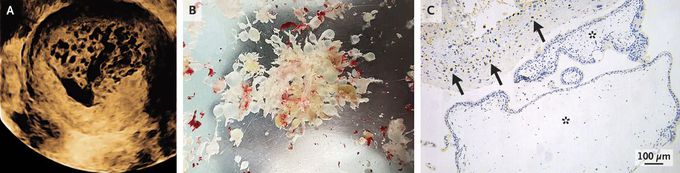
A 32-year-old woman who had a history of two pregnancies and deliveries presented to the gynecology clinic with a 20-day history of vaginal bleeding. Ten weeks before presentation, amenorrhea had developed, and a home pregnancy test had been positive 2 weeks before presentation. On physical examination, the fundal height was consistent with a pregnancy at 12 weeks’ gestation. The serum human chorionic gonadotropin (hCG) level was 222,634 IU per liter (reference value, <5). Transvaginal ultrasonography showed an intrauterine mass with numerous cystic spaces that created a “snowstorm” pattern (Panel A). Owing to concern about a hydatidiform mole, dilation and curettage was performed. Diffusely hydropic chorionic villi without any associated fetal parts were removed from the uterus (Panel B). Immunohistochemical analysis of a histopathological specimen showed expression of p57 — a maternally expressed gene product — in uterine tissue (Panel C, arrows) but not in the villi (asterisks). This finding confirmed the diagnosis of a complete hydatidiform mole, a type of gestational trophoblastic disease that results from an aberrant fertilization in which all chromosomes are paternal. Six weeks after surgery, the patient was doing well. The hCG level was undetectable at that time and remained so 1 year later.
