


Migrating Emboli in Branch Retinal Artery Occlusion
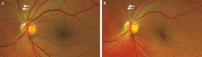
A 53-year-old man presented to the emergency department with sudden-onset, painless vision loss in the inferior visual field of the left eye. He had a history of an aortic aneurysm and had recently undergone multiple complex aortic-arch reconstructions. His postoperative course had been complicated by graft thrombosis, for which he had been taking apixaban and aspirin. On ophthalmologic examination, the visual acuity was 20/20 in the right eye and 20/25 in the left eye. An inferior defect was found on visual-field testing of the left eye. A funduscopic examination showed multiple filling defects in the superior division of the central retinal artery in the left eye, a finding consistent with platelet–fibrin emboli (Panel A, arrows). Trace surrounding macular edema without retinal whitening was also present. A diagnosis of branch retinal artery occlusion due to emboli from the aortic-arch graft thrombosis was made. Therapeutic anticoagulation was continued, and the patient’s visual deficit quickly improved. On repeat funduscopy 24 hours after presentation, the previously visualized emboli were absent, presumably as a result of migration (Panel B, arrows). The patient was discharged home and continued to receive therapeutic anticoagulation. At the 2-week follow-up, his visual acuity and findings on visual-field testing and funduscopic examination had normalized.

https://www.facebook.com/PrimalTRTOfficial/Acute Abdomen Differential DiagnosisFlexiLeafhttps://www.facebook.com/StableGripSafetyBarOfficial/https://www.facebook.com/HumeHealthBodyPodOfficial/Pulsar Vexline Review 2026 - Legit Or Scam Trading Platform? Fact Check
