


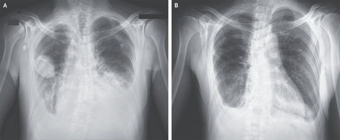
Phantom Tumor of the Lung
A 50-year-old man with a history of mitral-valve prolapse and of Hodgkin’s lymphoma 20 years earlier that was treated with chemotherapy and radiation therapy presented to the emergency department with a 5-day history of dyspnea. Physical examination showed jugular venous distention, diminished breath sounds at the lung bases, and a holosystolic murmur at the cardiac apex. A radiograph of the chest showed indistinct pulmonary vascularity, a dense retrocardiac opacity, and pleural effusions on both sides. A sharply demarcated, lenticular lesion was shown in the right middle lung field (Panel A, anterior–posterior view). An echocardiogram showed an ejection fraction of 45% and severe mitral regurgitation. Treatment with diuretics and vasodilators was initiated. A follow-up chest radiograph 3 days after presentation showed complete resolution of the right lung opacity, as well as reduced pulmonary edema (Panel B, posterior–anterior view). A diagnosis of phantom tumor of the lung was made. Phantom tumors of the lung, also known as vanishing tumors or pseudotumors, are loculated interlobar pleural effusions that occur most commonly in the minor fissure, as in this case. The resolution of phantom tumors with diuresis distinguishes them from true masses. Surgical coronary-artery revascularization and mitral-valve replacement were performed. At 1-month follow-up, the patient was doing well.
