

Bilothorax
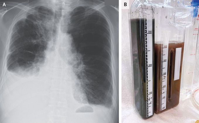
A 77-year-old man with metastatic lung adenocarcinoma presented to the emergency department with a 2-week history of dyspnea. Examination was notable for reduced breath sounds at the right lung base. A chest radiograph showed a pleural effusion on the right side (Panel A). An abdominal ultrasound image showed previously known liver metastases and perihepatic fluid, as well as new intrahepatic dilatation of the biliary ducts. Laboratory studies showed a serum total bilirubin level of 2.5 mg per deciliter (42.8 μmol per liter) (reference range, 0.3 to 1.2 mg per deciliter [5.1 to 20.5 μmol per liter]) and a direct bilirubin level of 2.0 mg per deciliter (34.2 μmol per liter) (reference range, 0.03 to 0.4 mg per deciliter [0.5 to 6.8 μmol per liter]). A chest tube was placed, and the color of the drained pleural fluid was olive brown that gradually changed to black (Panel B). Pleural-fluid studies showed an exudate with a total bilirubin level of 8.2 mg per deciliter (140.2 μmol per liter; reference value, 0) and a direct bilirubin level of 7.5 mg per deciliter (128.2 μmol per liter; reference value, 0). The pleural-fluid triglyceride level was normal, and cultures and cytologic studies were negative. A diagnosis of bilothorax was made. Bilothorax occurs when bile flows into the pleural space. In this case, the mechanism was thought to be diaphragmatic defects caused by hepatic metastases. Two weeks after presentation, the patient died from progressive liver failure.
I was diagnosed as a Hepatitis B carrier in 2015, with early signs of liver fibrosis. At first, antiviral medications helped control the virus but over time, resistance developed, and the effectiveness faded. I began to lose hope. In 2021, I discovered NaturePath Herbal Clinic despite my skepticism, I decided to give their herbal treatment a try.To my surprise, after just six months, my blood tests came back negative for the virus.It was nothing short of life-changing.I never expected such incredible results from a natural treatment. But it not only cleared the virus it restored my hope, my health, and my peace of mind.If you or someone you know is battling Hepatitis B, I truly encourage you to explore the natural healing path offered by NaturePath Herbal Clinic. It gave me a second chance and it might do the same for you.www.naturepathherbalclinic.com info@naturepathherbalclinic.com


