MEDizzy
MEDizzy
DR.MOHAMMED IRFAN SHEIKH
DR.MOHAMMED IRFAN SHEIKHabout 1 year ago
Calcium Polystyrene Sulfonate–Related Colonic Necrosis

Calcium Polystyrene Sulfonate–Related Colonic Necrosis

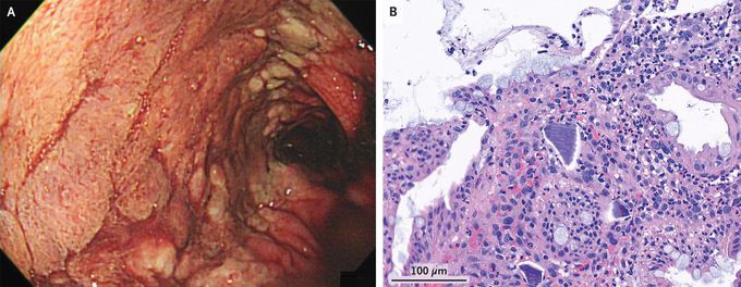
A 58-year-old woman who was hospitalized for acute-on-chronic kidney injury had sudden-onset bloody stools. At the time of admission, she had had hyperkalemia, and calcium polystyrene sulfonate — a cation-exchange resin that binds potassium in the colon — had been administered multiple times per day throughout the first 6 days of hospitalization. On hospital day 6, large-volume, painless hematochezia developed, along with a substantial decrease in the hemoglobin level. A colonoscopy revealed a continuous ulcer extending from the splenic flexure to the rectum (Panel A). There was no active bleeding. A biopsy of the colonic mucosa showed epithelial injury and basophilic calcium polystyrene sulfonate crystals in a necroinflammatory background (Panel B). A diagnosis of calcium polystyrene sulfonate–related colonic necrosis was made. In rare cases, gastrointestinal injury may occur with the use of calcium polystyrene sulfonate or its analogue, sodium polystyrene sulfonate. The true frequency and underlying mechanism are not known. The colon is the most frequently involved site. The patient’s hematochezia resolved with cessation of the culprit medication and bowel rest. She was discharged on hospital day 45 with improved renal function and a normal potassium level.

3
Other commentsSign in to post comments. You don't have an account? Sign up now!
Related posts
Foot X-ray AnatomyColonic adenocarcinomaColonic adenocarcinomaAnatomy of liverAbdominal aortic aneurysmImage of trachea blocked by chewing gum### Anatomy of the Heart: Summary The heart is a muscular organ responsible for pumping blood throughout the body, supplying oxygen and nutrients while removing waste products. It is located in the thoracic cavity, between the lungs, and is roughly the size of a fist. #### 1. **Structure:** - **Chambers:** The heart has four chambers: - **Right Atrium:** Receives deoxygenated blood from the body via the superior and inferior vena cavae. - **Right Ventricle:** Pumps deoxygenated blood to the lungs through the pulmonary arteries. - **Left Atrium:** Receives oxygenated blood from the lungs via the pulmonary veins. - **Left Ventricle:** Pumps oxygenated blood to the rest of the body through the aorta. - **Valves:** The heart contains four main valves that ensure unidirectional blood flow: - **Tricuspid Valve:** Located between the right atrium and right ventricle. - **Pulmonary Valve:** Located between the right ventricle and pulmonary arteries. - **Mitral (Bicuspid) Valve:** Located between the left atrium and left ventricle. - **Aortic Valve:** Located between the left ventricle and the aorta. #### 2. **Wall Layers:** - **Epicardium:** The outer layer, also known as the visceral pericardium. - **Myocardium:** The thick, muscular middle layer responsible for contraction. - **Endocardium:** The inner layer that lines the chambers and valves. #### 3. **Blood Supply:** - The heart receives its blood supply from the coronary arteries, which branch off the aorta. The **right coronary artery** supplies the right side of the heart, while the **left coronary artery** branches into the left anterior descending and circumflex arteries, supplying the left side. #### 4. **Electrical System:** - The heart's rhythmic contractions are controlled by its electrical conduction system, which includes: - **Sinoatrial (SA) Node:** The natural pacemaker located in the right atrium. - **Atrioventricular (AV) Node:** Receives impulses from the SA node and transmits them to the ventricles. - **Bundle of His and Purkinje Fibers:** Distribute the electrical impulse throughout the ventricles, leading to contraction. #### 5. **Function:** - The heart functions in two primary circulatory loops: - **Pulmonary Circulation:** Moves deoxygenated blood from the right side of the heart to the lungs for oxygenation. - **Systemic Circulation:** Distributes oxygenated blood from the left side of the heart to the rest of the body. The anatomy of the heart is crucial for its role in maintaining effective circulation and overall cardiovascular health. Understanding its structure helps in diagnosing and treating various heart conditions.Acute tubular necrosisIschaemic Acute tubular necrosisToxic Acute tubular necrosis

Recent MCQs


Show more MCQs

Recent flashcard sets


Show more flashcards