


Retinoblastoma
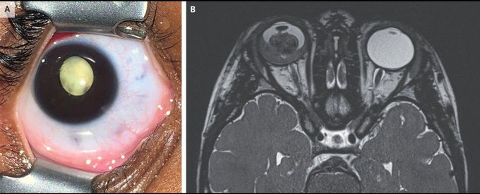
A 3-year-old girl was brought to the emergency department with a 2-month history of a white pupil and a 1-day history of redness and pain in the right eye. An eye examination performed while the patient was under anesthesia showed leukocoria (Panel A), as well as iris neovascularization and a white, nodular mass in the posterior chamber. The left eye was normal. B-scan ultrasonography showed tumor calcification and vitreous seeding in the affected eye. A diagnosis of retinoblastoma was made. Magnetic resonance imaging of the head showed retinal and choroidal invasion without extraocular extension (Panel B). Given the high-risk clinical features, the eye was enucleated the next day. Histopathological examination confirmed the diagnosis. Genetic testing did not show a germline mutation in the tumor suppressor gene RB1, and therefore the tumor was attributed to a somatic, nonheritable mutation. Leukocoria requires urgent evaluation by an ophthalmologist in order to quickly identify life-threatening causes. When detected early, retinoblastoma can be treated with focal therapy that can salvage vision and the eye. When diagnosis is delayed, however, the condition is often treated with enucleation and systemic chemotherapy to prevent life-threatening metastasis. After enucleation, the patient underwent six cycles of chemotherapy. There was no evidence of recurrent disease on subsequent examinations.
