

Concomitant Urinary Triple Phosphate and Cystine Crystals
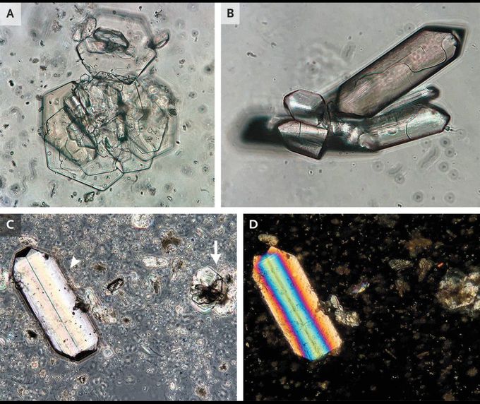
A 37-year-old man with cystinuria was seen in the nephrology clinic for routine follow-up. He had received a diagnosis of cystinuria at 1 year of age, when he had presented with nephrolithiasis and pyelonephritis; subsequent workup had shown elevated 24-hour cystine urinary excretion and homozygous variants in the gene SLC3A1. Since then, he had been treated with urinary alkalinization, high fluid intake, and tiopronin. At the current presentation, he was asymptomatic. The serum creatinine level was normal, and a urinalysis showed a pH of 7.0, with no hematuria or bacteriuria. Microscopic examination of a spun urine specimen showed hexagonal cystine crystals (Panel A) and rhombohedral triple phosphate — or struvite — crystals (Panel B). In the same field of view elsewhere in the specimen (Panel C), triple phosphate crystals of a different shape (arrowhead) and cystine crystals (arrow) were seen. Under polarized light, these triple phosphate crystals showed polychromatic properties, and the cystine crystals showed moderate polarizing properties (Panel D). Triple phosphate crystals are associated with urease-producing organisms. The patient had been treated for urinary tract infection 2 weeks before this presentation, but the causative organism had been unknown. At follow-up 1 month later, the patient had no recurrent symptoms of urinary tract infection.

