MEDizzy
MEDizzy
DR.MOHAMMED IRFAN SHEIKH
DR.MOHAMMED IRFAN SHEIKHabout 1 year ago
Isolated Persistent Left Superior Vena Cava

Isolated Persistent Left Superior Vena Cava

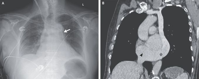
A 79-year-old man with a history of urinary retention that had been treated with an indwelling urinary catheter was admitted to the intensive care unit for septic shock. On hospital day 1, ultrasonographic guidance was used to insert a hemodialysis catheter into the right internal jugular vein to initiate renal replacement therapy for oliguric acute kidney injury. A chest radiograph obtained after the procedure showed the catheter crossing the midline at the level of the manubrium and entering the left mediastinum (Panel A, arrow). Arterial placement of the catheter was initially suspected, although blood gas analysis was consistent with venous placement. Retrospective review of recent computed tomographic images of the chest (Panel B) showed an isolated persistent left superior vena cava coursing along the left mediastinum and subsequently connecting with a large coronary sinus (asterisk) that drained into the right atrium (arrowhead). A persistent left superior vena cava, which is often detected incidentally, is the most common anomaly of the thoracic venous system. It typically occurs simultaneously with a right superior vena cava, which was not found in this patient. Culture of a urine specimen from this patient grew Escherichia coli. After treatment with vasopressors, antibiotic agents, and renal replacement therapy, the septic shock and acute kidney injury resolved. The hemodialysis catheter was removed without complication.

2
Other commentsSign in to post comments. You don't have an account? Sign up now!
Related posts
Long BonesFlat Bones
Stomach
Foot X-ray AnatomyAnatomy of liverAbdominal aortic aneurysmImage of trachea blocked by chewing gum### Anatomy of the Heart: Summary The heart is a muscular organ responsible for pumping blood throughout the body, supplying oxygen and nutrients while removing waste products. It is located in the thoracic cavity, between the lungs, and is roughly the size of a fist. #### 1. **Structure:** - **Chambers:** The heart has four chambers: - **Right Atrium:** Receives deoxygenated blood from the body via the superior and inferior vena cavae. - **Right Ventricle:** Pumps deoxygenated blood to the lungs through the pulmonary arteries. - **Left Atrium:** Receives oxygenated blood from the lungs via the pulmonary veins. - **Left Ventricle:** Pumps oxygenated blood to the rest of the body through the aorta. - **Valves:** The heart contains four main valves that ensure unidirectional blood flow: - **Tricuspid Valve:** Located between the right atrium and right ventricle. - **Pulmonary Valve:** Located between the right ventricle and pulmonary arteries. - **Mitral (Bicuspid) Valve:** Located between the left atrium and left ventricle. - **Aortic Valve:** Located between the left ventricle and the aorta. #### 2. **Wall Layers:** - **Epicardium:** The outer layer, also known as the visceral pericardium. - **Myocardium:** The thick, muscular middle layer responsible for contraction. - **Endocardium:** The inner layer that lines the chambers and valves. #### 3. **Blood Supply:** - The heart receives its blood supply from the coronary arteries, which branch off the aorta. The **right coronary artery** supplies the right side of the heart, while the **left coronary artery** branches into the left anterior descending and circumflex arteries, supplying the left side. #### 4. **Electrical System:** - The heart's rhythmic contractions are controlled by its electrical conduction system, which includes: - **Sinoatrial (SA) Node:** The natural pacemaker located in the right atrium. - **Atrioventricular (AV) Node:** Receives impulses from the SA node and transmits them to the ventricles. - **Bundle of His and Purkinje Fibers:** Distribute the electrical impulse throughout the ventricles, leading to contraction. #### 5. **Function:** - The heart functions in two primary circulatory loops: - **Pulmonary Circulation:** Moves deoxygenated blood from the right side of the heart to the lungs for oxygenation. - **Systemic Circulation:** Distributes oxygenated blood from the left side of the heart to the rest of the body. The anatomy of the heart is crucial for its role in maintaining effective circulation and overall cardiovascular health. Understanding its structure helps in diagnosing and treating various heart conditions.

Recent MCQs


Show more MCQs

Recent flashcard sets


Show more flashcards