


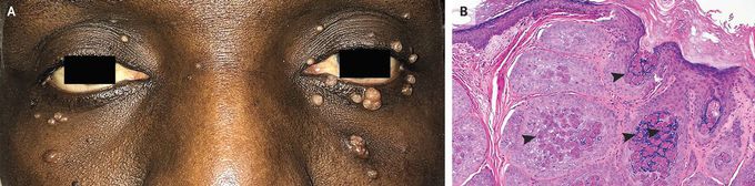
Molluscum Contagiosum
A 60-year-old man with a history of human immunodeficiency virus (HIV) infection presented to the ophthalmology clinic with lesions on his eyelids and face that had increased in number during the preceding 4 months. His CD4 cell count was 20 per cubic millimeter (reference range, 500 to 1500) and his HIV viral load was 120,000 copies per milliliter. Numerous dome-shaped, flesh-colored papules with central umbilication were observed on both the eyelids and the cheeks (Panel A). Incision and curettage of all the lesions were performed, followed by the application of prophylactic antibiotic ointment. Histopathological assessment of the lesions showed coarse, basophilic, keratohyaline granules with large intracytoplasmic, eosinophilic inclusion bodies (Panel B, arrowheads), confirming a diagnosis of molluscum contagiosum. Molluscum contagiosum is caused by a poxvirus and leads to chronic, localized infection of the skin that can occur anywhere on the body. Patients with cellular immunodeficiency are at risk for more severe, treatment-resistant infection. The differential diagnosis for molluscum contagiosum includes cryptococcosis, histoplasmosis, basal-cell carcinoma, and warts. The patient was referred for HIV treatment. At a follow-up visit 6 months later, he had had no recurrence of the lesions.

Living with HIV was one of the hardest experiences of my life. The fatigue, the emotional toll, and the uncertainty about the future weighed on me every single day. I had tried many treatments and medications, but nothing seemed to restore my health or energy the way I hoped.Out of both hope and desperation, I came across NaturePath Herbal Clinic. At first, I was skeptical but something about their natural approach and the powerful stories I read gave me the courage to try one more time.I began their herbal treatment program, and within a few weeks, I noticed small but meaningful changes more energy, better sleep, and a stronger immune system. Over the months, those improvements only grew. Today, I can truly say my life has changed. I feel healthier, more balanced, and finally in control of my well-being again.This isn’t just a testimony it’s a heartfelt recommendation to anyone living with HIV or any chronic condition. Don’t give up hope. I’m so grateful I gave NaturePath Herbal Clinic a chance. Visit their website to learn more: www.naturepathherbalclinic.com Email: info@naturepathherbalclinic.com
