


Tinea Capitis Due to Microsporum canis
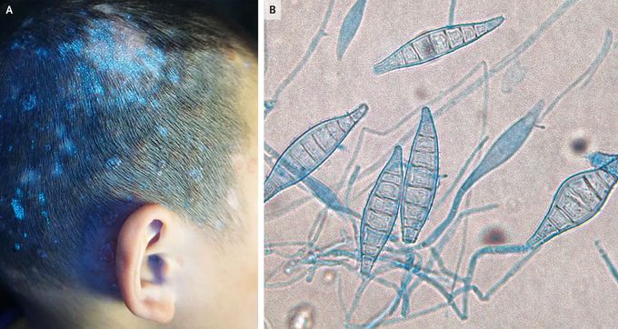
A 6-year-old boy presented to the dermatology clinic with a 3-month history of rash on his face and scalp, with associated hair loss. On physical examination, he had multiple scaly, erythematous plaques on his scalp, neck, and face as well as patches of alopecia and bilateral postauricular lymphadenopathy. Areas of bright green fluorescence were observed on his scalp on Wood’s lamp examination (Panel A). After preparation of a hair sample with potassium hydroxide, microscopic examination showed hyphae and spores on the outside of hair shafts, indicating an ectothrix infection. Microsporum canis was identified on molecular testing of scalp scrapings, and direct microscopic evaluation of fungal cultures showed septate hyphae and macroconidia (Panel B). M. canis is a zoophilic fungus that is often acquired from domestic animals. The patient’s parents reported no pets at home but mentioned that the patient may have had exposure to animals while staying in the countryside several months earlier. Treatment with oral and topical terbinafine was initiated, and after 6 weeks of therapy, the skin lesions had resolved. The parents were contacted 3 and 6 months after the completion of therapy, and they confirmed that there had been no recurrence of the rash.
