


Ocular Findings in the Sturge–Weber Syndrome
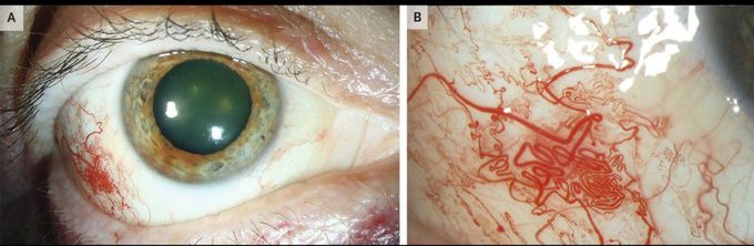
A 62-year-old man with the Sturge–Weber syndrome was referred to the glaucoma clinic for assessment of high intraocular pressure in his right eye. Physical examination showed a port-wine birthmark over the maxillary area on the right side of his face. The intraocular pressure was 25 mm Hg (reference range, 11 to 21) in the right eye and was normal in the left eye. Examination of the anterior segment of the right eye showed a moderate cataract as well as focal dilatation and tortuosity of the conjunctival and episcleral vasculature (Panels A and B). The Sturge–Weber syndrome is a congenital vascular disorder involving capillary and venous malformations in the brain, skin, and eyes and is strongly associated with glaucoma. The intraocular pressure is determined by the rate at which aqueous humor is produced in the eye relative to the rate at which drainage occurs through the episcleral venous plexus. Downstream vascular malformations in the Sturge–Weber syndrome can increase episcleral venous pressure, causing dilatation of the ocular surface vessels and increasing intraocular pressure. The patient was treated with a topical carbonic anhydrase inhibitor to suppress the production of aqueous humor and received a miotic medication to increase outflow through the trabecular meshwork. At follow-up 1 month later, his intraocular pressure had normalized.
