MEDizzy
MEDizzy
DR.MOHAMMED IRFAN SHEIKH
DR.MOHAMMED IRFAN SHEIKHabout 1 year ago
Kayser–Fleischer Rings in Wilson’s Disease

Kayser–Fleischer Rings in Wilson’s Disease

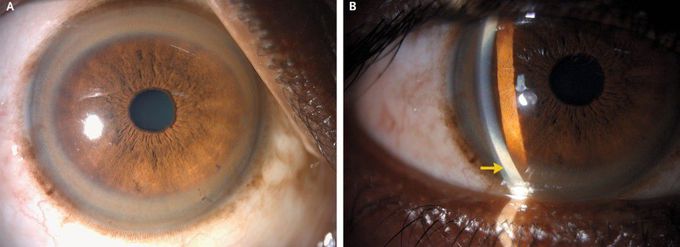
A 47-year-old man with no known medical history presented to the department of internal medicine with a 2-month history of abdominal distention and melena. He did not drink alcohol or take any medications or supplements. Physical examination was notable for ascites, pedal edema, gynecomastia, postural tremor, and mild cognitive impairment. During the physical examination, pigmented rings were observed in both corneas (Panel A), and copper deposition was observed in Descemet’s membrane on slit-lamp examination (Panel B, arrow). Both findings were consistent with Kayser–Fleischer rings. Laboratory studies showed mild elevations in serum levels of aminotransferases and total bilirubin, a prolonged international normalized ratio, and a normal alkaline phosphatase level. The serum ceruloplasmin level was 3.1 mg per deciliter (reference range, 14 to 40), the serum copper level was 5 μmol per liter (reference range, 10 to 22), and the 24-hour urinary copper excretion was 382 μg (reference range, 10 to 30). Genetic testing revealed a compound heterozygous mutation in ATP7B, the gene encoding a copper-transporting ATPase, and a diagnosis of cirrhosis due to Wilson’s disease was made. Treatment with trientine and zinc for copper chelation was started, and the patient was listed for liver transplantation.

3
Other commentsSign in to post comments. You don't have an account? Sign up now!
Related posts
Stomach
Foot X-ray AnatomyAnatomy of liverAbdominal aortic aneurysmImage of trachea blocked by chewing gum### Anatomy of the Heart: Summary The heart is a muscular organ responsible for pumping blood throughout the body, supplying oxygen and nutrients while removing waste products. It is located in the thoracic cavity, between the lungs, and is roughly the size of a fist. #### 1. **Structure:** - **Chambers:** The heart has four chambers: - **Right Atrium:** Receives deoxygenated blood from the body via the superior and inferior vena cavae. - **Right Ventricle:** Pumps deoxygenated blood to the lungs through the pulmonary arteries. - **Left Atrium:** Receives oxygenated blood from the lungs via the pulmonary veins. - **Left Ventricle:** Pumps oxygenated blood to the rest of the body through the aorta. - **Valves:** The heart contains four main valves that ensure unidirectional blood flow: - **Tricuspid Valve:** Located between the right atrium and right ventricle. - **Pulmonary Valve:** Located between the right ventricle and pulmonary arteries. - **Mitral (Bicuspid) Valve:** Located between the left atrium and left ventricle. - **Aortic Valve:** Located between the left ventricle and the aorta. #### 2. **Wall Layers:** - **Epicardium:** The outer layer, also known as the visceral pericardium. - **Myocardium:** The thick, muscular middle layer responsible for contraction. - **Endocardium:** The inner layer that lines the chambers and valves. #### 3. **Blood Supply:** - The heart receives its blood supply from the coronary arteries, which branch off the aorta. The **right coronary artery** supplies the right side of the heart, while the **left coronary artery** branches into the left anterior descending and circumflex arteries, supplying the left side. #### 4. **Electrical System:** - The heart's rhythmic contractions are controlled by its electrical conduction system, which includes: - **Sinoatrial (SA) Node:** The natural pacemaker located in the right atrium. - **Atrioventricular (AV) Node:** Receives impulses from the SA node and transmits them to the ventricles. - **Bundle of His and Purkinje Fibers:** Distribute the electrical impulse throughout the ventricles, leading to contraction. #### 5. **Function:** - The heart functions in two primary circulatory loops: - **Pulmonary Circulation:** Moves deoxygenated blood from the right side of the heart to the lungs for oxygenation. - **Systemic Circulation:** Distributes oxygenated blood from the left side of the heart to the rest of the body. The anatomy of the heart is crucial for its role in maintaining effective circulation and overall cardiovascular health. Understanding its structure helps in diagnosing and treating various heart conditions.Fuction of ears: Your ears have two main functions: hearing and balance. Hearing: When sound waves enter your ear canal, your tympanic membrane (eardrum) vibrates. This vibration passes on to three tiny bones (ossicles) in your middle ear. The ossicles amplify and transmit these sound waves to your inner ear. Once the sound waves reach your inner ear, tiny hair cells called stereocilia transform the vibrations into electrical energy and send it along nerve fibers to your brain. Balance: Your inner ear contains semicircular canals filled with fluid and hair-like sensors. When you move your head, the fluid inside these loop-shaped canals sloshes around and moves the hairs. The hairs transmit this information along the vestibular nerve to your brain. Finally, your brain sends signals to your muscles to help you stay balanced.Ear injury Cuts, fractures and blunt force trauma can cause ear injury. If damage is severe, surgery may be necessary to address the problem. This may include surgery to preserve hearing or cosmetic surgery to improve the appearance of your ear.Ear tumors Ear tumors may be benign (noncancerous) or malignant (cancerous). Types of noncancerous ear tumors include keloids, sebaceous cysts, osteomas and exostoses (bone growths). Noncancerous ear tumors usually require surgical removal. Cancers that can affect your ears include melanoma, basal cell carcinoma and squamous cell carcinoma. Treatment for these conditions depends on several factors, including the type and stage of cancer, and whether or not it has spread to other parts of your body.

Recent MCQs


Show more MCQs

Recent flashcard sets


Show more flashcards