


Abdominopelvic Actinomycosis
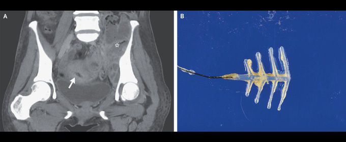
A 54-year-old woman presented to the emergency department with a 2-month history of fevers and weight loss and a 3-week history of abdominal pain and difficulty walking. Her temperature was 38.5°C. Examination was notable for a tender mass in the left lower quadrant of the abdomen. Laboratory studies showed a white-cell count of 30,420 per cubic millimeter (reference range, 3300 to 8600). Contrast-enhanced computed tomography (CT) revealed an intrauterine device (IUD) (Panel A, arrow) surrounded by multiple abscesses (asterisk) throughout the abdomen and pelvis that extended into the left iliopsoas muscle and left hip joint. The patient underwent CT-guided drainage of the abscesses, washout of the left hip joint, and removal of the IUD, which was coated in sulfur-colored granules (Panel B). Gram’s staining of fluid samples revealed branching, filamentous, gram-positive rods. Actinomyces israelii was grown on culture. The patient reported that the IUD had been inserted more than two decades before presentation and had not been replaced since that time. After a prolonged course of intravenous antibiotics followed by oral antibiotics, the patient’s abdominal and hip pain abated.
