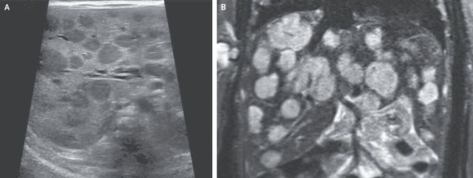


Infantile Hepatic Hemangiomas
A 16-day-old girl who had not yet returned to her birth weight was brought to the emergency department with lethargy. She had tachypnea and marked hepatomegaly with a hepatic bruit, as well as eight small hemangiomas on the scalp, thorax, abdomen, and limbs that had been noted within days after birth. Abdominal ultrasonography showed numerous rounded hypoechoic lesions in the liver (Panel A). Whole-body magnetic resonance imaging showed contrast-enhancing hepatic lesions, as large as 20 mm in diameter, that were hyperintense on T2-weighted images (Panel B), a finding consistent with infantile hepatic hemangiomas. Moderate cardiomegaly was also noted. Laboratory studies showed a thyrotropin level of 25.5 mIU per liter (reference range, 0.50 to 8.50) and a reverse triiodothyronine level of greater than 3696 pmol per liter (reference range, 140 to 540). Complications of infantile hepatic hemangiomas include high-output cardiac failure, consumptive hypothyroidism, and bleeding. These vascular lesions typically have rapid growth followed by a phase of slow involution, sometimes over a period of years. Treatment with propranolol, furosemide, and high-dose levothyroxine was initiated. The liver lesions gradually reduced in size, and after 9 months, treatment was stopped successfully.
I was diagnosed as a Hepatitis B carrier in 2015, with early signs of liver fibrosis. At first, antiviral medications helped control the virus but over time, resistance developed, and the effectiveness faded. I began to lose hope. In 2021, I discovered NaturePath Herbal Clinic despite my skepticism, I decided to give their herbal treatment a try.To my surprise, after just six months, my blood tests came back negative for the virus.It was nothing short of life-changing.I never expected such incredible results from a natural treatment. But it not only cleared the virus it restored my hope, my health, and my peace of mind.If you or someone you know is battling Hepatitis B, I truly encourage you to explore the natural healing path offered by NaturePath Herbal Clinic. It gave me a second chance and it might do the same for you.www.naturepathherbalclinic.com info@naturepathherbalclinic.com


