


Pulmonary Mucormycosis
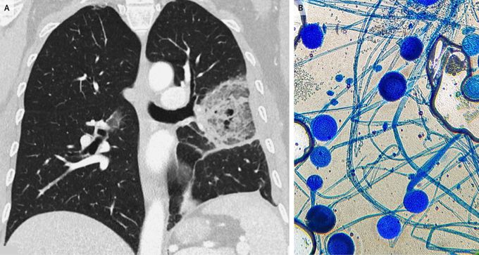
A 51-year-old woman with a history of acute lymphoblastic leukemia presented to the emergency department with pleuritic pain in the left side of the chest that had persisted for 3 days; she had no fever or cough. She had undergone bone marrow transplantation 3 months previously, and graft-versus-host disease had developed, for which she had been receiving treatment with cyclosporine and high doses of glucocorticoids. Physical examination was notable for decreased breath sounds on the left side. Laboratory studies revealed a glycated hemoglobin level of 9.6% (reference range, 4.0 to 5.6). A computed tomographic scan of the chest showed an area of ground-glass opacity surrounded by a denser ring of consolidation (Panel A), a finding referred to as the reversed halo sign. Bronchoalveolar lavage was performed, and on microscopic examination with lactophenol cotton-blue staining, a culture of the specimen showed mycotic structures consistent with rhizopus (Panel B). A diagnosis of pulmonary mucormycosis was made. Mucormycosis is an invasive fungal infection that is encountered primarily in patients with a high degree of immunosuppression. The reversed halo sign on imaging has been described in other infectious and noninfectious conditions; however, in severely immunocompromised patients, this sign is suggestive of infection by an invasive fungal pathogen, as was observed in our patient. Treatment with liposomal amphotericin B was initiated, and surgery was planned; however, the patient died 15 days after presentation.
