


Spontaneous Hyphema
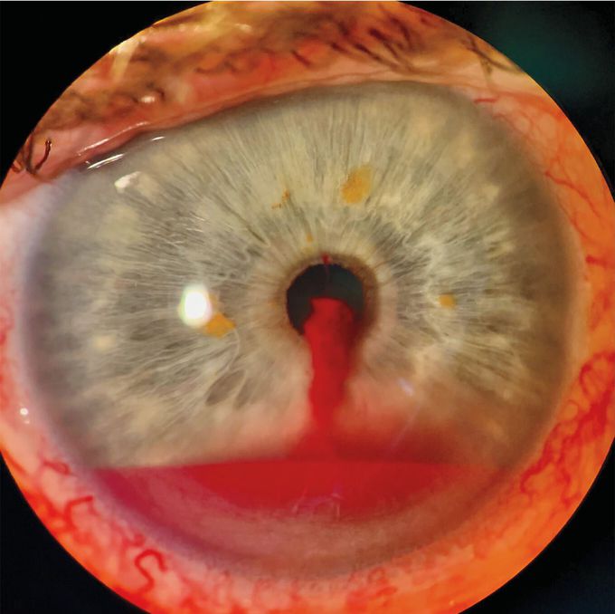
A 72-year-old man with well-controlled type 2 diabetes presented to an ophthalmology clinic with a 4-hour history of discomfort and blurry vision in his right eye. He reported no eye trauma or regular use of anticoagulants. Eye examination showed conjunctival injection of the right eye with a visual acuity of 20/30. Extraocular movements and pupillary responses were intact. On slit-lamp biomicroscopy, an enlarging pool of blood was seen in the anterior chamber, a condition known as hyphema. The blood originated from an actively bleeding iris microhemangioma at the superior pupillary margin (see video). The examination was otherwise notable for mild nonproliferative diabetic retinopathy and normal intraocular pressures in both eyes. A diagnosis of spontaneous hyphema due to a bleeding iris microhemangioma was made. Although hyphema most often occurs in the context of ocular trauma, it can occur spontaneously as a result of vascular lesions of the iris, intraocular cancers, or ocular ischemia. Conservative treatment with bed rest and head elevation was recommended, and topical atropine and topical dexamethasone were prescribed to minimize ciliary spasm and intraocular inflammation, respectively. At a 10-day follow-up visit, the patient’s symptoms had abated, and laser photocoagulation of the microhemangioma was deferred. Parth R. Shah, F.R.A.N.Z.C.O. Sydney, Australia

Brain Blood SupplyAnatomy of Oral CavityNeck Muscles
