MEDizzy
MEDizzy
Sheeza Basharat
Sheeza Basharatabout 2 years ago
Aortic Stenosis

Aortic Stenosis

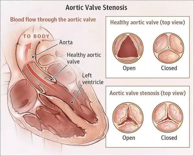
Aortic stenosis is a narrowing of the aortic valve, the outflow valve of the heart between the left ventricle (main pumping chamber) and the aorta (major blood vessel carrying blood to the body).The aortic valve normally functions as a 1-way valve that prevents blood from leaking back into the heart. When the aortic valve narrows, the heart has to work harder to pump blood into the aorta ✔Symptoms of aortic stenosis include chest pressure, lightheadedness or fainting, breathlessness, and/or fatigue. They indicate that the extra work required to open the valve has overcome the heart’s ability to work normally. ✔Medications are usually not effective in treating aortic stenosis as this is a mechanical problem, and valve replacement is the only proven therapy to improve symptoms and prolong life.

Source: https://www.instagram.com/p/CgSMy5AsQfA/?igshid=MjU0Y2ZlMmY=
4
Other commentsSign in to post comments. You don't have an account? Sign up now!
Related posts
Atrial DepolarisationST SegmentQRS ComplexNephrotic Syndrome### Anatomy of the Heart: Summary The heart is a muscular organ responsible for pumping blood throughout the body, supplying oxygen and nutrients while removing waste products. It is located in the thoracic cavity, between the lungs, and is roughly the size of a fist. #### 1. **Structure:** - **Chambers:** The heart has four chambers: - **Right Atrium:** Receives deoxygenated blood from the body via the superior and inferior vena cavae. - **Right Ventricle:** Pumps deoxygenated blood to the lungs through the pulmonary arteries. - **Left Atrium:** Receives oxygenated blood from the lungs via the pulmonary veins. - **Left Ventricle:** Pumps oxygenated blood to the rest of the body through the aorta. - **Valves:** The heart contains four main valves that ensure unidirectional blood flow: - **Tricuspid Valve:** Located between the right atrium and right ventricle. - **Pulmonary Valve:** Located between the right ventricle and pulmonary arteries. - **Mitral (Bicuspid) Valve:** Located between the left atrium and left ventricle. - **Aortic Valve:** Located between the left ventricle and the aorta. #### 2. **Wall Layers:** - **Epicardium:** The outer layer, also known as the visceral pericardium. - **Myocardium:** The thick, muscular middle layer responsible for contraction. - **Endocardium:** The inner layer that lines the chambers and valves. #### 3. **Blood Supply:** - The heart receives its blood supply from the coronary arteries, which branch off the aorta. The **right coronary artery** supplies the right side of the heart, while the **left coronary artery** branches into the left anterior descending and circumflex arteries, supplying the left side. #### 4. **Electrical System:** - The heart's rhythmic contractions are controlled by its electrical conduction system, which includes: - **Sinoatrial (SA) Node:** The natural pacemaker located in the right atrium. - **Atrioventricular (AV) Node:** Receives impulses from the SA node and transmits them to the ventricles. - **Bundle of His and Purkinje Fibers:** Distribute the electrical impulse throughout the ventricles, leading to contraction. #### 5. **Function:** - The heart functions in two primary circulatory loops: - **Pulmonary Circulation:** Moves deoxygenated blood from the right side of the heart to the lungs for oxygenation. - **Systemic Circulation:** Distributes oxygenated blood from the left side of the heart to the rest of the body. The anatomy of the heart is crucial for its role in maintaining effective circulation and overall cardiovascular health. Understanding its structure helps in diagnosing and treating various heart conditions.Tinzaprin CalculationsCephalosporinsGlycopeptidesSickle Cell Disease VIISeizures I

Recent MCQs


Show more MCQs

Recent flashcard sets


Show more flashcards