


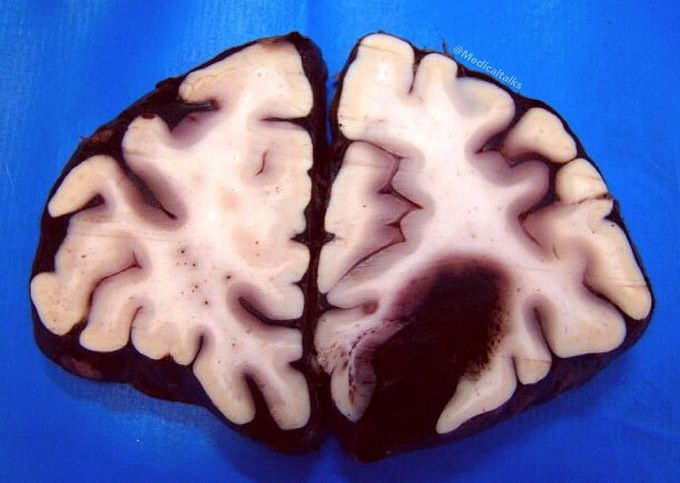
Cerebral hemorrhage
Gross specimen of a brain, frontal section, showing a cerebral hemorrhage (cerebrovascular accident) of the left hemisphere in a patient with hypertension!! There is a large hematoma with mass effect. Hemorrhages involving the basal ganglia area (the putamen in particular) tend to be non-traumatic and caused by hypertension, which damages and weakens the small penetrating branches of the major cerebral arteries. The resulting spontaneous deep intracerebral bleeding caused a mass effect with midline shift, often with secondary edema, may disrupt or compress adjacent brain tissue, leading the neurological dysfunction. Substantial displacement of brain parenchyma may cause elevation of intracranial pressure and potentially fatal herniation syndromes. Nontraumatic intracerebral hemorrhage most commonly results from hypertensive damage to blood vessel walls (eg, hypertension, eclampsia, drug abuse), but it also may be due to autoregulatory dysfunction with excessive cerebral blood flow (eg, reperfusion injury, hemorrhagic transformation), rupture of an aneurysm or arteriovenous malformation, or arteriopathy (eg, cerebral amyloid angiopathy, moyamoya). Nonsurgical management is considered for patients with minimal neurological deficits or with intracerebral hemorrhage volumes less than 10 mL. Surgery is preserved for patients with cerebellar hemorrhage greater than 3 cm, for patients with intracerebral hemorrhage associated with a structural vascular lesion, and for young patients with lobar hemorrhage.
