
Dr.rasoulover 7 years ago
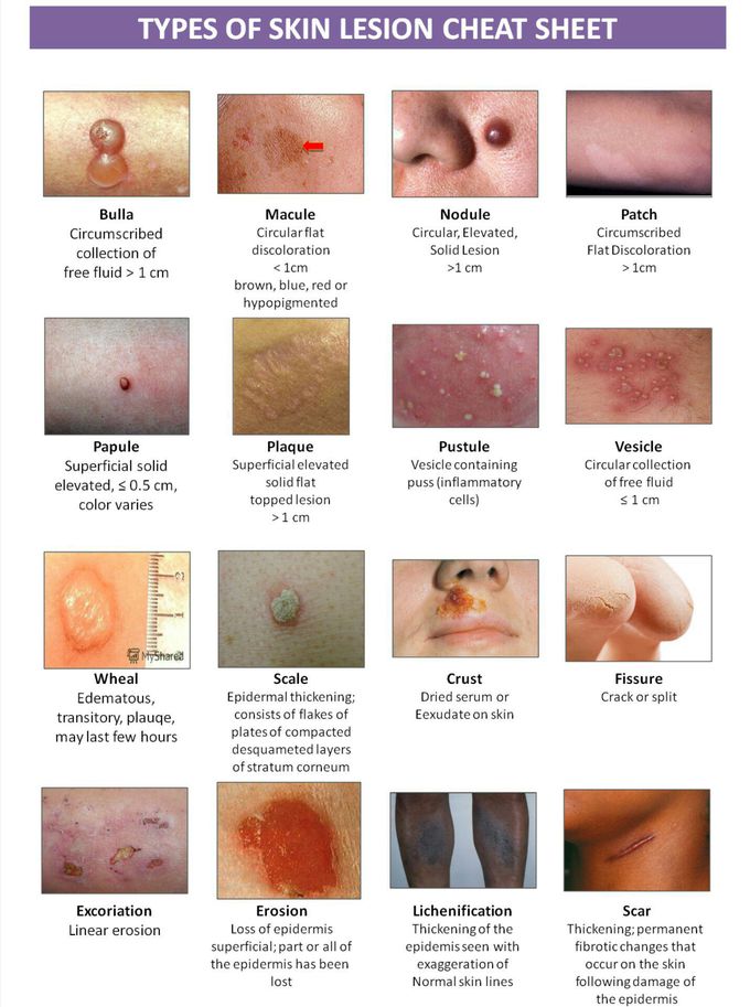
Tutorial A: The Primary Lesions Primary lesions are physical changes in the skin considered to be caused directly by the disease process. Types of primary lesions are rarely specific to a single disease entity. 1.MACULE A macule is a change in the color of the skin. It is flat, if you were to close your eyes and run your fingers over the surface of a purely macular lesion, you could not detect it. A macule greater than 1 cm. may be referred to as a patch. 2. PAPULE A papule is a solid raised lesion that has distinct borders and is less than 1 cm in diameter. Papules may have a variety of shapes in profile (domed, flat-topped, umbilicated) and may be associated with secondary features such as crusts or scales. 3. NODULE A nodule is a raised solid lesion more than 1 cm. and may be in the epidermis, dermis, or subcutaneous tissue. 4. TUMOR A tumor is a solid mass of the skin or subcutaneous tissue; it is larger than a nodule. (Please bear in mind this definition does not at all mean that the lesion is a neoplasm.) 5. PLAQUE A plaque is a solid, raised, flat-topped lesion greater than 1 cm. in diameter. It is analogous to the geological formation, the plateau. 6. VESICLE Vesicles are raised lesions less than 1 cm. in diameter that are filled with clear fluid. 7. BULLAE Bullae are circumscribed fluid-filled lesions that are greater than 1 cm. in diameter. 8. PUSTULE Pustules are circumscribed elevated lesions that contain pus. They are most commonly infected (as in folliculitis) but may be sterile (as in pustular psoriasis) 9. WHEAL A wheal is an area of edema in the upper epidermis. 10. BURROW Burrows are linear lesions produced by infestation of the skin and formation of tunnels (e.g., with infestation by the scabitic mite or by cutaneous larva migrans). 11. TELANGIECTASIA Telangiectasia are the permanent dilatation of superficial blood vessels in the skin and may occur as isolated phenomena or as part of a generalized disorder, such as ataxia telangiectasia. Tutorial B: The Secondary Lesions Secondary lesions may evolve from primary lesions, or may be caused by external forces such as scratching, trauma, infection, or the healing process. The distinction between a primary and secondary lesion is not always clear. 12. SCALE Scale consists of flakes or plates that represent compacted desquamated layers of stratum corneum. Desquamation occurs when there are peeling sheets of scale following acute injury to the skin. 13. CRUST Crusting is the result of the drying of plasma or exudate on the skin. Please remember that crusting is different from scaling. The two terms refer to different phenomena and are not interchangeable. One can usually be distinguished from the other by appearance alone. 14. ATROPHY Atrophy is thinning or absence of the epidermis or subcutaneous fat. 15. LICHENIFICATION "Lichenification" refers to a thickening of the epidermis seen with exaggeration of normal skin lines. It is usually due to chronic rubbing or scratching of an area. 16. EROSION Erosions are slightly depressed areas of skin in which part or all of the epidermis has been lost. 17. EXCORIATION Excoriations are traumatized or abraded skin caused by scratching or rubbing. 18. FISSURE A fissure is linear cleavage of skin which extends into the dermis. 19. ULCERATION Ulcerations occur when there is necrosis of the epidermis and dermis and sometimes of the underlying subcutaneous tissue. 20. SCAR Scars are the permanent fibrotic changes that occur on the skin following damage to the dermis. Scars may have secondary pigment characteristics. 21. ESCHAR An eschar is a hard, usually darkened, plaque covering an ulcer implying extensive tissue necrosis, infarcts or gangrene. 22. KELOIDS Keloids are an exaggerated connective tissue response of injured skin that extend beyond the edges of the original wound. 23. PETECHIAE, PURPURA, AND ECCHYMOSES Three terms that refer to bleeding that occurs in the skin are petechiae, purpura, and ecchymoses. Generally, the term "petechiae" refers to smaller lesions. "Purpura" and "ecchymoses" are terms that refer to larger lesions. In certain situations purpura may be palpable. In all situations, petechiae, ecchymoses, and purpura do not blanch when pressed. If there is any question, press on the lesions carefully with a glass slide. Don't break the slide or cut the patient. Tutorial C: Patterns and Distribution Not only is the appearance of lesions important, but the pattern and distribution on the skin is as well. 24. ANNULAR Annular lesions are seen in a ring shape. Tinea corporis, erythema migrans (the lesion associated with lyme disease), and granuloma annulare are three common examples. 25. DISCRETE Discrete lesions tend to remain separate. This is a helpful descriptive term but has little specific diagnostic significance. 26. CLUSTERED Clustered lesions are those that are grouped together. They are commonly seen in herpes simplex or with insect bites, for example. 27. CONFLUENT Confluent lesions tend to run together. 28. DERMATOMAL, ZOSTERIFORM Dermatomal, zosteriform lesions follow a dermatome. The lesions of varicella zoster (also known as shingles) are the classic example, but there are other lesions which may assume the same pattern. 29. ECZEMATOID Eczematoid lesions are inflamed with a tendency toward clustering, oozing, or crusting. 30. FOLLICULAR It is sometimes helpful to determine if lesions specifically involve the hair follicle. 31. GUTTATE Guttate lesions look as though someone took a dropper and dropped this lesion on the skin. Guttate lesions are characteristic of one form of psoriasis, though that is not the only example. 32. IRIS OR TARGET LESIONS Iris lesions are also known as target lesions and are a series of concentric rings. These have a dark or blistered center. These lesions are frequently seen with erythema multiforme but not exclusively so. 33. KOEBNER PHENOMENON The Koebner phenomenon, also called the isomorphic response, refers to the appearance of lesions along a site of injury. This phenomenon is seen in a variety of conditions; for example, lichen planus, warts, molluscum contagiosum, psoriasis, lichen nitidus, and the systemic form of juvenile rheumatoid arthritis. 34. LINEAR LESIONS Linear lesions occur in a line or band-like configuration. This descriptive term may apply to a wide variety of disorders. (One should be certain that the lesions are not following a dermatome.) 35. MULTIFORM Patients with multiform lesions have lesions of a variety of shapes. 36. RETICULAR Reticular or net-like lesions can be seen in a variety of circumstances; e.g., very commonly in newborns (or even grown children and adults) as cutis marmorata, or with livedo reticularis. The former fades as the skin is warmed the latter becomes more florid. 37. SERPIGINOUS Serpiginous lesions wander as though following the track of a snake. 38. UNIVERSALIS Universalis refers to a widespread disorder that affects the entire skin. 39. SCARLATINIFORM Scarlatiniform rashes have the pattern of scarlet fever. The patient with a scarlatiniform rash has innumerable small red papules that are widely and diffusely distributed. Note that the term scarlatiniform does not mean that the patient has scarlet fever, although by definition all patients with scarlet fever have a scarlatiniform rash. Patients with a variety of other conditions such as Kawasaki disease, viral infections, or drug reactions may have rashes with the same pattern. 40. STRAWBERRY TONGUE Patients with scarlet fever, Kawasaki disease or other conditions may develop a distinctive appearance of their tongues. Because of its resemblance to the well-known berry, the appearance is called "strawberry tongue." 41. MORBILLIFORM The term "morbilliform" means that the patient has a rash that looks like measles. Patients with measles will have the rash but patients with Kawasaki disease, drug reactions, or other conditions may also have a morbilliform rash. The rash consists of macular lesions that are red and are usually 2-10 mm in diameter but may be confluent in places. 42. SATELLITE LESIONS The term is commonly used to describe a portion of the rash of cutaneous candidiasis in which a beefy red plaque may be found surrounded by numerous, smaller red macules located adjacent to the body of the main lesions. 43. PATTERNS OF INTENTIONAL OR UNINTENTIONAL INJURY One important category of skin lesions involve the form that skin lesions may take in cases of child abuse or other intentional injury (bite marks, slap marks, strap marks, burns, etc.) or in cases of unintentional injury. Abrasions are traumatically caused erosions.
Other commentsSign in to post comments. You don't have an account? Sign up now!