


SM
Syed Mohammad Hasanabout 1 year ago
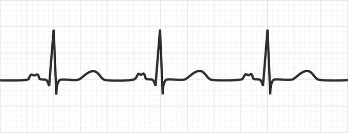
Bifid P Waves AKA P Mitrale
The P wave on ECG represents atrial depolarization. It is a first positive deflection with smooth contour. Changes in the normal morphology of P wave may indicate some underlying pathology. Bifid or notched P wave on ECG indicates left atrial enlargement. The notched appearance of P wave is attributed to longer time taken by left atrium as compared to the right atrium. The duration of P wave is usually longer than 120 ms. The bifid P waves can be appreciated in lead II. Reference: https://litfl.com/p-wave-ecg-library/ Image via: https://medschool.co/tests/ecg-basics/the-p-wave
Other commentsSign in to post comments. You don't have an account? Sign up now!
Related posts
ECG Review IECG Review IIWe use cookies to enhance your browsing experience, serve personalized ads or content, and analyze our traffic. By clicking "Accept All", you consent to our use of cookies.
Best Anabolic Steroids Natural Muscle Boosters: A 2025 ReviewElectrocardiogram (ECG) - Normal sinus rhythm (NSR): Clinical Nursing CarePediatric ECGECG Rhythms
Best Anabolic Steroids Natural Muscle Boosters: A 2025 ReviewElectrocardiogram (ECG) - Normal sinus rhythm (NSR): Clinical Nursing CarePediatric ECGECG Rhythms
