


Mishal Shan7 months ago
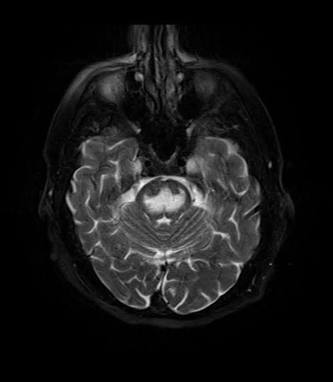
Central Pontine Myelinosis
The given image is a T2 weighted MRI scan of the brain which shows hyperintensity in the pons secondary to pontine myelinosis. This is most often an iatrogen condition resulting from rapid correction of hyponatremia. The pathophysiology behind the condition is as follows. During states of hyponatremia, the brain cells adapt to decrease intracellular osmolality so the cells still remain isotonic to the extracellular environment. If blood levels of sodium rapidly increase, the ECF becomes hypertonic without allowing brain cells sufficient time to adapt. This leads to osmotic imbalance and cells continue to lose water and are unable to function. https://en.m.wikipedia.org/wiki/Central_pontine_myelinolysis
Other commentsSign in to post comments. You don't have an account? Sign up now!
Related posts
