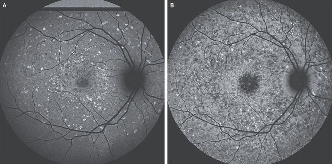


Stargardt Juvenile Macular Degeneration
A 13-year-old boy was referred to the ophthalmology clinic with enlarging blind spots in the central vision in both eyes. He had no family history of eye problems. At presentation, the visual acuity was 20/40 in the right eye and 20/50 in the left eye. Fundus examination of both eyes showed widespread, yellow, pisciform flecks. Short-wavelength autofluorescence imaging showed diffuse hyperautofluorescent lesions suggestive of bisretinoid accumulation and patchy central areas of atrophy of the retinal pigment epithelium with peripapillary sparing (Panel A). These findings are consistent with a diagnosis of Stargardt juvenile macular degeneration (also called Stargardt’s disease), an autosomal recessive disorder caused by mutations in the gene encoding ATP-binding cassette subfamily A member 4 (ABCA4). Dysfunction of the ABCA4 transporter leads to the accumulation of toxic bisretinoids with consequent degeneration of the retinal pigment epithelium and photoreceptors. Vision loss typically begins during childhood and progresses. The diagnosis was confirmed with sequencing of ABCA4. The patient was advised to avoid vitamin A supplementation, which may accelerate bisretinoid accumulation. At 6-year follow-up, visual acuity was 20/70 in the right eye and 20/50 in the left eye, with worsening central scotomata. Autofluorescence imaging showed progressive atrophy in the previously identified areas of pisciform flecks with the maintenance of peripapillary sparing

