Double Superior Venae Cavae!
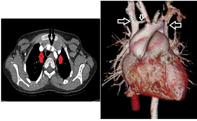
During normal development, the superior vena cava develops from right anterior cardinal and right common cardinal veins. Persistence of the left anterior cardinal vein results in a left SVC. This left SVC drains into the right atrium through the coronary sinus. In the case of double vena cava, the anastomosis between the anterior cardinal veins which forms the left brachiocephalic vein is also small or absent. In above shown figures the double venae cavae can be seen. Axial CECT image shows two superior venae cavae (marked red arrow) and the intercommunicating vein (marked black arrow). In figure 2, 3D Ct angiographic image shows double superior venae cavae (marked horizontal arrows) with the venous inter communication (marked vertical arrow). Images and their description via: https://www.imagejournals.org/articles/double-superior-vena-cava-115.html