Lung Herniation after Cardiac Surgery
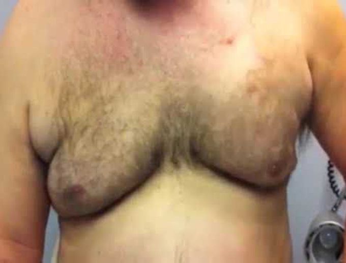
A 53-year-old man presented with left pectoral swelling. He had undergone minimally invasive, robot-assisted multivessel coronary revascularization 15 months earlier. He noted that the swelling became more prominent on straining or coughing. On physical examination, a soft area of swelling was noted that extended from the left third to the left fifth anterior ribs; the swelling became more evident during a Valsalva maneuver and during coughing (Video). Computed tomographic (CT) images of the chest (Panel A, axial plane; and Panel B, coronal plane) show a lung herniation through two intercostal levels that was caused by muscular defects in the chest wall. CT images of the chest obtained during a Valsalva maneuver show an increase in the amount of herniation (Panel C, axial plane; and Panel D, coronal plane). Lung herniation is a rare complication of minimally invasive cardiothoracic surgery.