


Endoscopic-assisted post. interhemi retrocallosal transfalcine approach for falcotentorial ...
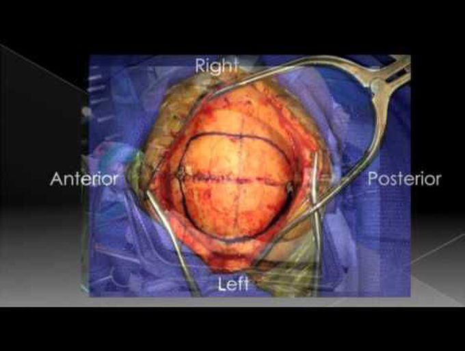
Falcotentorial meningiomas are rare tumors of the pineal region that arise from the dural folds where the falx and tentorium meet and are often intimately related to the vein of Galen and straight sinus. These lesions often present with signs and symptoms related to hydrocephalus and brainstem compression. Surgical resection of falcotentorial meningiomas remains the definitive treatment, with a variety of surgical approaches used to resect these lesions. The choice of approach depends on several factors, including the size and location of the vein of Galen complex relative to the tumor. Falcotentorial meningiomas can be technically challenging to remove with significant risk of morbidity because of the close proximity to and occasional invasion of the vein of Galen and straight sinus. In this operative video, the authors demonstrate an illustrative step-by-step technique for endoscopic-assisted microsurgical resection of a falcotentorial meningioma using the posterior interhemispheric retrocallosal transfalcine approach for a superiorly positioned falcotentorial meningioma. The surgical nuances are discussed, including the surgical anatomy, gravity-assisted interhemispheric approach in the lateral position, retrocallosal dissection, transfalcine exposure, tumor removal, and preservation of the vein of Galen complex. In summary, the posterior interhemispheric retrocallosal transfalcine approach is a useful surgical strategy for select superiorly positioned falcotentorial meningiomas. James K. Liu, MD, and Michael A. Cohen, MD Department of Neurological Surgery, Center for Skull Base and Pituitary Surgery, Neurological Institute of New Jersey, Rutgers New Jersey Medical School, Newark, New Jersey
