


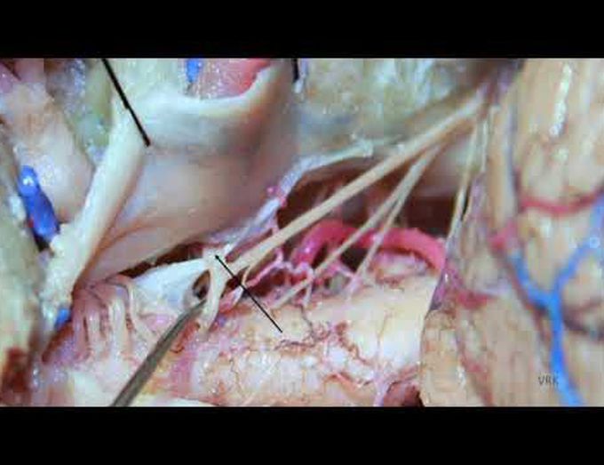
Far lateral approach for microsurgical ligation of C1 dAVF: surgical anatomy and technical nuances
Department of Neurosurgery, Cleveland Clinic, Cleveland, Ohio This video demonstrates the diagnosis and surgical ligation of a C1 dural arteriovenous fistula via a far lateral, transcondylar approach. The patient’s dural arteriovenous fistula was identified by MRI signal changes in the spinal cord and a cerebrospinal angiogram demonstrating an abnormal hypertrophied early venous drainage pattern suggestive of a C1 vessel origin. Indocyanine green was used to verify surgical treatment of the fistula intraoperatively. A postoperative angiogram and MR image demonstrate fistula occlusion and resolution of the spinal cord edema. Anatomic details and technical nuances of the approach are demonstrated. Varun R. Kshettry, MD, Nina Z. Moore, MD, and Mark Bain, MD
