


Internal Carotid Artery in the Middle Ear
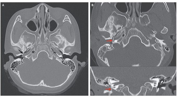
A 6-year-old girl presented with a 1-year history of pulsatile tinnitus in her right ear. Pure-tone audiometry showed normal hearing. Otoscopy showed a whitish pulsatile mass behind the tympanic membrane in the anteroinferior quadrant of the middle-ear cavity (see video). Computed tomography (CT) of the temporal bone revealed a vessel coursing through the hypotympanum (Panel A, arrow) and entering the carotid canal through a dehiscence in the carotid plate — a finding consistent with an aberrant right internal carotid artery. Normal structures in the left ear are shown for comparison, including the normal location of the internal carotid artery at this level within the carotid canal and an intact carotid plate. An aberrant internal carotid artery in the middle ear is a rare vascular anomaly. It is essentially a collateral pathway that traverses the middle ear, involving the communication of an enlarged inferior tympanic artery with an enlarged caroticotympanic artery, and can occur when the cervical internal carotid artery is underdeveloped or has regressed during embryogenesis. An aberrant internal carotid artery can mimic other middle-ear lesions, so it is important that imaging be performed before any surgical intervention in order to avoid bleeding complications that may result from misdiagnosis. The patient underwent surgical correction (Panel B, arrows; the upper panel shows an axial CT scan, and the lower panel a coronal CT scan). At 13 months of follow-up, otoscopy showed a healthy tympanic drum, and the patient reported no remaining symptoms.
Video link. https://youtu.be/MqOK_h24R5k
