


Embryology of Kidney system , The development of kidney
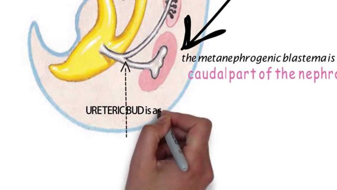
This illustration video deals with the development of kidney sysytes, the development of Metaneohros.The facts have been presented in a way that makes clear sense. In just 13 min whole topic has been dealt with good details. Very use full, beautiful and highly comprehensive illustration with a lot of high yield facts and figures. Metanephros[edit] During the fifth week of gestation, the mesonephric duct develops an outpouching, the ureteric bud, near its attachment to the cloaca. This bud, also called the metanephrogenic diverticulum, grows posteriorly and towards the head of the embryo. The elongated stalk of the ureteric bud, called the metanephric duct, later forms the ureter. As the cranial end of the bud extends into the intermediate mesoderm, it undergoes a series of branchings to form the collecting duct system of the kidney. It also forms the major and minor calyces and the renal pelvis. The portion of undifferentiated intermediate mesoderm in contact with the tips of the branching ureteric bud is known as the metanephrogenic blastema. Signals released from the ureteric bud induce the differentiation of the metanephrogenic blastema into the renal tubules. As the renal tubules grow, they come into contact and join with connecting tubules of the collecting duct system, forming a continuous passage for flow from the renal tubule to the collecting duct. Simultaneously, precursors of vascular endothelial cells begin to take their position at the tips of the renal tubules. These cells differentiate into the cells of the definitive glomerulus. In humans, all of the branches of the ureteric bud and the nephronic units have been formed by 32 to 36 weeks of gestation. However, these structures are not yet mature, and will continue to mature after birth. Once matured, humans have an estimated two million nephrons (approximately 1,000,000 per kidney) but this number is highly variable ranging widely from approximately 300,000 to over 2 million per kidney The development of the kidney proceeds through a series of successive phases, each marked by the development of a more advanced kidney: the pronephros, mesonephros, and metanephros.[1] The pronephros is the most immature form of kidney, while the metanephros is most developed. The metanephros persists as the definitive adult kidney. Metanephros Definitive Kidney permanent kidney begin to develop early in the fifth week and start to function approximately 4 weeks later two sources The metanephric diverticulum The metanephrogenic blastema The metanephric diverticulum is an outgrowth from the mesonephric the metanephrogenic blastema is derived from the caudal part of the nephrogenic cord The stalk of the metanephric diverticulum becomes the urete cranial portion of the diverticulum undergoes repetitive branching events differentiate into the collecting tubules of the metanephros ). By day 32, each ureteric bud penetrates a portion of the sacral intermediate mesoderm called the metanephric blastema The first four generations of tubules enlarge to form the major calices the second four generations form the minor calices The end of each arched collecting tubule induces clusters of mesenchymal cells in the metanephrogenic blastema to form small metanephric vesicles (Fig. 12-7A). These vesicles elongate and become the metanephros, or permanent kidney Urine formation continues throughout fetal life The lobulation usually disappears during infancy as the nephrons increase and grow each kidney containing 400,000 to 2,000,000 nephrons A uriniferous tubule consists of two embryologically different parts A nephron derived from the metanephrogenic blastema A collecting tubule derived from the metanephric diverticulum By the middle of the 6th week, the developing metanephros consists of two lobes separated by a sulcus. By the end of the 16th week, 14 to 16 lobes have formed ( When the ureteric bud first contacts the metanephric blastema, its tip expands to form an initial ampulla Nephrogenesis is complete by birth in humans. Potter sequence Steps in renogenesis reciprocal induction Derivatives of the metanephric blastema • Podocytes covering glomerular capillaries • Epithelial cells lining Bowman’s capsule • Proximal convoluted tubules • Descending thick limbs of the loops of Henle • Thin limbs of the loops of Henle • Ascending thick limbs of the loop of Henle • Distal convoluted tubules Derivatives of the ureteric bud • Collecting tubules and ducts • Minor and major calyces • Ureters Nephroblastoma (Wilms Tumor
