

Mitral Annular Calcification
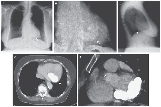
An 81-year-old woman was transferred to the medical service with cardiac failure after surgery for oral cancer. A diagnostic workup indicated perioperative overhydration and pulmonary embolism as causative factors for her symptoms. Chest radiography revealed a large, dense area of calcification overlying the heart, consistent with mitral annular calcification. Calcification was semicircular in the location of the mitral valve, as seen on the preoperative posterior–anterior view (Panel A, arrowhead; Panel B, arrow, shows an enlarged view) and a lateral view (Panel C, arrowhead). Computed tomography revealed calcification of the posterolateral mitral-valve annulus on the axial view (Panel D, arrowhead) and the short-axis view (Panel E). Transthoracic echocardiography revealed moderate mitral regurgitation and stenosis, left ventricular diastolic dysfunction, and a left ventricular ejection fraction of 69%. Mitral annular calcification is often detected on routine chest radiographs in elderly patients and is considered to be a degenerative condition that is usually unrelated to clinical symptoms. When mitral annular calcification is massive, it can lead to valvular dysfunction (as in this patient), typically resulting in complete heart block, mitral regurgitation, or less often, mitral stenosis. Mitral annular calcification may be related to diabetes, hypertension, hyperlipidemia, and secondary hyperparathyroidism from renal failure. During her hospitalization, the patient was conservatively treated with oral heart medication. No surgery was performed, and the patient was lost to follow-up.

